 Figure 2. Cross section of a reserve fang of a seasnake. Note functional hollow.
An examination of a fang from any of the above families will show a non-functional anterior groove, which is no more than an enamel-sealed seam. Shine (1991: 15) labels this external groove as such.
The continual reference to grooved front fangs, and the incorrect inference that the venom flows down this groove, probably originated by mistake as a typo error, but has persisted through the years. Imagine a scientist presenting a paper on the evolutionary development of snake fangs. He explains how the ancestral state was an open anterior canal, or groove, and illustrates this with a several drawings of an open canal, an elapid or hydrophid fang (the more primitive state in front-fanged snakes) and a viper's fang. As he points to the most obvious closed seam in an elapid or hydrophid fang he says, "See how it was grooved." The possible typo error was the transposing of it was with it's.
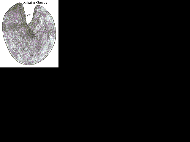 Figure 1. Cross-section of rear fang of colubrid. Note functional groove! VC= venom canal.
I believe 'groove' is another word that is best consigned to the scrap heap when describing the fangs in front-fanged snakes. The only fangs with a functional groove (ectoglyphous) for envenomation are those (Figure 1) in back-fanged snakes (opisthoglyphous). All others have an enclosed venom canal (Figure 2) and are effectively hollow (endoglyphous).
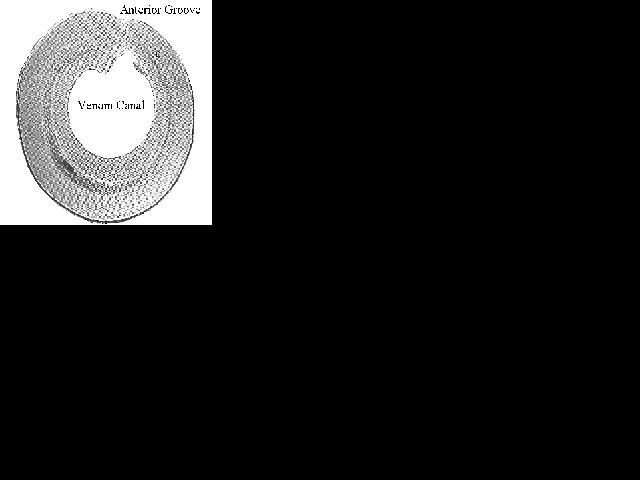
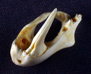
Have a look at the following figure after Limpus (1987) showing the cross section of a seasnake fang. This is almost identical to the fang in elapids and vipers. Limpus states, "The outer enamel layer of the tooth is continuous across the front face of this tubular fang."
Figure 2. Cross section of a reserve fang of a seasnake. Note functional hollow.
Compare the following drawing of a fang from a viper, the puff adder (Bitis arietans), taken from Parker & Grandison's (1977) work, Snakes - a Natural History page 41, with the illustration on page 15 in Shine (1991). They are all but identical!
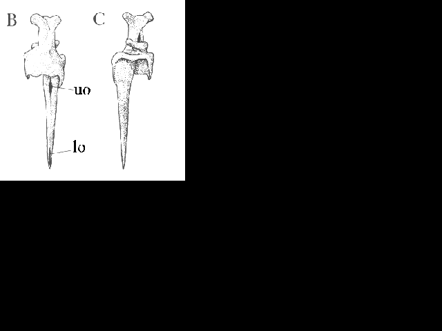 Figure 3. Anterior (B) and posterior (C) views of maxilla. uo upper orifice; lo lower orifice
An article in the July edition of the Hawkesbury Herpetologist prompted me to include this piece in our newsletter. In that article it said,
"Australian snake fangs are not hollow like many overseas species, but rather a fang with a grooved edge at the rear."
How the myth persists! Considering his statement I can only assume the author has never had the opportunity to examine an Australian snake's fang. Do not make the same mistake. Those writers not exercising enough care perpetuate many a myth. I should know. I have made mistakes previously. I just hope, as the years slip by, I make fewer than I did in my younger days.
In elapids and seasnakes these teeth generally have an enclosed hollow. This may have been an open groove in its ancestral state, but not today. (For illustrations of elapid fangs see Shine 1991: Australian Snakes a Natural History. Pg 15 & 189 and Mirtschin & Davis (revised) 1991: Dangerous Snakes of Australia page 39. For seasnake fangs see Limpus 1987: pages 194-95 In Toxic Plants and Animals a Guide for Australia).
In rear-fanged colubrids several conditions occur in the following order of frequency. 1) Enlarged teeth with an open anterior groove (proectoglyphous); 2) enlarged teeth with an open lateral groove (pleuroglyphous); and 3) no fangs per se, only slightly enlarged teeth almost indistinguishable from the adjacent teeth.
I am unaware of any Australian elapid with the condition described in the Hawkesbury Herpetologist.
The primary difference between the fangs in elapids/hydrophids and those in vipers is the length (Figure 4) and the degree they can be moved. The latter are generally much longer and more movable (solenoglyphous), whereas the former are small and fixed (proteroglyphous).
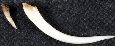
Figure 4. Comparison of length in Aussie Mulga Snake fang (left) and African Gaboon Viper (right).
To describe any endoglyph as having a grooved venom canal is pedantic in the extreme.
Remember though! In the fangs of all front-fanged snakes there is the non-functional, enamel-sealed, anterior seam. It is an irrelevant groove (Figure 5).
Note: In Australia there are 136 known species plus a couple of undescribed taxa of front-fanged and 6 rear-fanged venomous snakes. In WA, 80 (56 elapids, 24 hydrophiids) and 4 respectively. This follows those listed in Cogger (1995) plus Boiga fusca, Acanthophis wellsi, Austrelaps labialis, A. ramsayi, Demansia calodera, D. reticulata, D. rufescens, Rhinoplocephalus bushi, Vermicella intermedia, V. multifasciata, V. snelli, V. vermiformis.